From Jonathan Lambert Science News
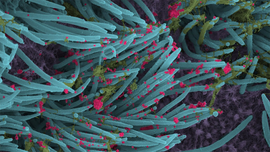
New closeup views of lung cells show just how prolifically the coronavirus that causes COVID-19 can replicate once it infiltrates the respiratory tract.
In the lab, pediatric pulmonologist Camille Ehre and colleagues at the University of North Carolina at Chapel Hill infected cells that line the airways in the lungs with SARS-CoV-2, waited 96 hours and then snapped scanning electron micrograph images of the virus-laden cells.
“Once a cell is infected, it is completely taken over by the virus, producing an astonishing number of viruses,” Ehre says. In a lab dish of about 1 million human cells, she says the viral load can skyrocket from about one thousand infectious viruses to 10 million in just two days. The new images were published September 3 in the New England Journal of Medicine.
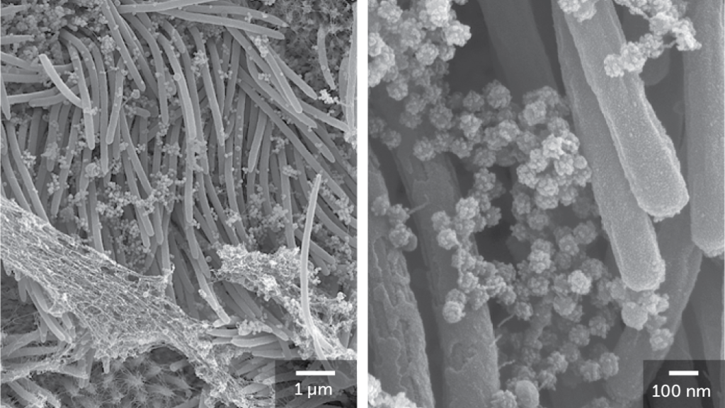
Cells that line the respiratory tract and their hairlike protrusions called cilia help clear airways of inhaled particles and pathogens. These types of cells are also specifically targeted by the coronavirus. Once infected, they churn out “astronomical numbers” of viral particles, Ehre says, potentially propelling the particles deeper into the lungs, which can cause pneumonia, or out into the air where they can infect others. Read more from Science News
Read other science related stories from News Without Politics