
How the cell binds the virus: Covid Virus under the helium ion microscope for the first time, Bielefeld University, Germany
Scientists at the Faculty of Physics have succeeded for the first time in imaging the SARS-CoV-2 coronavirus with a helium ion microscope.
Written content from by Bielefeld University via Phys
In contrast to the more conventional electron microscopy, the samples do not need a thin metal coating in helium ion microscopy.
This allows interactions between the coronaviruses and their host cell to be observed particularly clearly. The scientists have published their findings, obtained in collaboration with researchers from the Bielefeld University’s Medical School OWL and Justus Liebig University Giessen, in the Beilstein Journal of Nanotechnology.
The study shows that the helium ion microscope is suitable for imaging coronaviruses—so precisely that the interaction between virus and host cell can be observed,” says physicist Dr. Natalie Frese.
She is the lead author of the study and a researcher in the research group Physics of Supramolecular Systems and Surfaces at the Faculty of Physics.
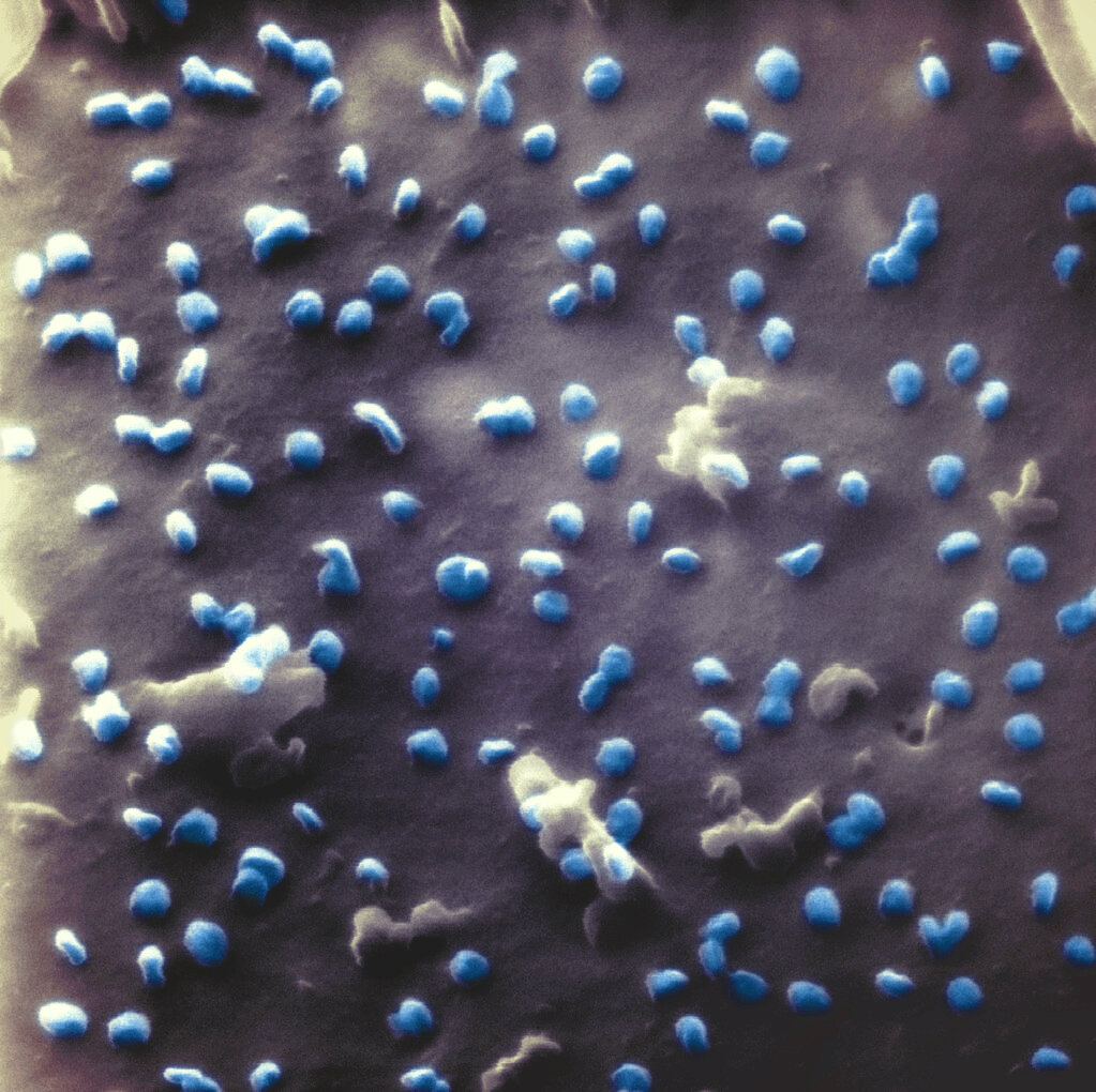
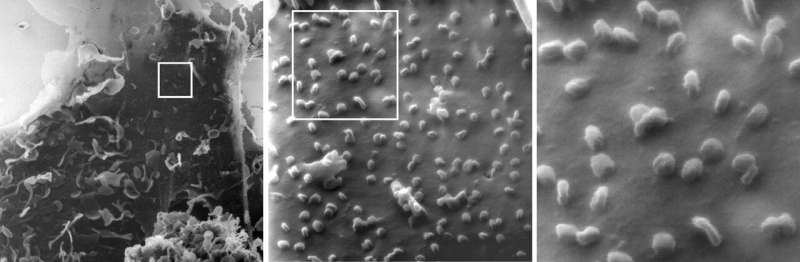
Coronaviruses are tiny—only about 100 nanometres in diameter, or 100 billionths of a meter. So far, mainly scanning electron microscopy (SEM) has been used to examine cells infected with the virus. With SEM, an electron beam scans the cell and provides an image of the surface structure of the cell occupied by viruses.
However, SEM has a disadvantage: the sample becomes electrostatically charged during the microscopy process. Because the charges are not dispersed from non-conductive samples, for example viruses or other biological organisms, the samples must be coated with an electrically conductive coating, such as a thin layer of gold.
“However, this conductive coating also changes the surface structure of the sample. Helium ion microscopy does not require a coating and therefore allows direct scanning,” says Professor Dr. Armin Gölzhäuser, who heads the research group Physics of Supramolecular Systems and Surfaces.
With the helium ion microscope, a beam of helium ions scans the surface of the sample. Helium ions are helium atoms that are each missing an electron—they are therefore positively charged. The ion beam also charges the sample electrostatically, but this can be compensated for by additionally irradiating the sample with electrons. Furthermore, the helium ion microscope has a higher resolution and a greater depth of field.
In their study, the scientists infected cells—artificially produced from the kidney tissue of a species of monkey—with SARS-CoV-2 and studied them in dead state under the microscope. “Our images provide a direct view of the 3-D surface of the coronavirus and the kidney cell—with a resolution in the range of a few nanometres,” says Frese. This enabled the researchers to visualize interactions between the viruses and the kidney cell.
Their study results indicate, for example, that helium ion microscopy can be used to observe whether individual coronaviruses are just lying on the cell or are bound to it. This is important in order to understand defense strategies against the virus: an infected cell can bind the viruses, which have already multiplied inside it, to its cell membrane on exit and thus prevent them from spreading further. Read more from Phys